Graphium
Corda (1837)
What isGraphium?
GenusGraphiumcomprises species of asexual, filamentous fungi belonging to the familyMicroascaceae, class Sordariomycetes, and subclass Hypocreomycetidae [1]. Sexual states ofGraphiumspecies occur inPseudallescheriaandPetriella, while species such asG. basitruncatumdon’t have a known sexual stage [2].
Where canGraphiumbe found?
Graphiumspp. can be found insoil, plant material, and woody substrate [1, 3]. It is rarely isolated as a contaminant. However, some species of the genus are capable of infecting humans. These species (including sexual states) belong to the generaPetriella,Pseudallescheria, andCeratocystis[1].
What doesGraphiumlook like?
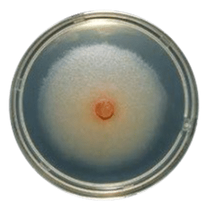
Graphium形成光灰色,羊毛柔软的殖民地t mature within seven days(Fig. 1). From the reverse, colonies are initially pale, becoming darker as they mature [1].

(Image source: Cruywagen, E. et al. (2010) [6])
Some sexually reproducing fungi can have several types of asexual states, and these are called synanamorphs. For example, sexually reproducingPseudallescheria boydiican have two types of anamorphic (sexual) states –Scedosporium apiospermum(scedosporiumsynanamorph) andGraphium eumorphum(Graphiumsynanamorph). Both synanamorphs do not always have to appear in an isolate. They can appear successfully at different stages, or both can be present, with one being located at the edges of a colony, as is sometimes observed inP. boydii[5].
What are health effects ofGraphium?
The most clinically important isolates ofGraphiumbelong to the genusScedosporium, which includes its asexual stages [1,5].Scedosporium apiospermum and Scedosporium prolificansare ubiquitous molds commonly found in soil, polluted waters, and sewage. They can cause a broad spectrum of clinical diseases, known as scedosporiosis, mostly in immunocompromised patients [5]. These fungi can colonize already damaged bronchia and lungs, particularly in cases of tuberculosis and cystic fibrosis [5]. The entry route is respiratory, and symptoms often include sinusitis, pneumonia, fever, cough, and coughing blood, followed by abnormal breathing sounds [5].
Infection can also follow the injury of skin and subcutaneous tissues, causing necrotic and ulcerated lesions. Infection can be localized or disseminated through the bloodstream to distant organs such as tendons, ligaments, bones, joints, the heart, or the brain [5].Pseudallescheria boydii, the sexual stage ofS. apiospermum, is also involved in infections. It can be recognized by the presence of a fruiting body (cleistothecia) [5].
Another clinically important species ofGraphiumisGraphium basitruncatum, reported as a causative agent of infections in immunocompromised patients [2, 3]. There has been one case of subcutaneous infection caused by this fungus in a heart transplant patient. The only symptom was a lesion in the palm since the patient didn’t have a fever or any sign of septicemia [3]. The other reported case of infection caused byGraphium basitruncatumwas persistent fungemia (presence of fungal cells in the blood) in a patient with acute leukemia. Symptoms included fever and skin lesions on the patient’s extremities, and the outcome was fatal [2].
Diagnosis ofGraphiumspecies is traditionally based on culture and morphological characterization, while molecular tools are still investigational [5]. Clinical specimens can be obtained by biopsy of infected tissue, aspiration from sinuses, or blood samples.Graphium物种可以被成功地种植在日常劳动atory media such as Sabouraud glucose agar, blood agar, and chocolate agar [3, 5], as well as on selective media such as modified Leonian agar that inhibits the growth of other filamentous fungi [5]. The incubation temperature is 25 °C to 35 °C (77 °F- 95°F) for allScedosporiumstrains, while some of them can grow at 37°C (98.6 °F) or 42°C (107.6 °F) [5]. The reported incubation temperature for the cultivation ofGraphium basitruncatumis 30°C (86 °F) to 37°C (98.6 °F) [3].
How to treat infections caused byGraphiumspecies?
While infections caused byS. apiospermum(P. boydii) can be treated with antifungal triazoles, infections caused byS. prolificansrarely respond to medical therapy without surgery and immunotherapy [5]. Since only two cases of human infection withGraphium basitruncatumhave been reported, there is no data referring to successful treatment.
References
- GraphiumRetreated from:https://drfungus.org/knowledge-base/graphium-species/
- Kumar, D. et al. (2007).Graphium basitruncatumFungemia in a Patient with Acute Leukemia. Journal od Clinical Microbiology. 45(5): 1644–1647.
- Fernández, A. L. et al. (2017). Subcutaneous infection byGraphium basitruncatumin a heart transplant patient. The Brazilian Journal of Infectious Diseases. 21 (6): 670-674
- Geldenhuis, M. M. et al. (2004). Identification and pathogenicity ofGraphiumandPesotumspecies from machete wounds onSchizolobium parahybumin Ecuador. Fungal diversity 15: 137-151
- Cortez, K. J. (2008). Infections Caused by Scedosporium spp. Clinical Microbiology Review. 21 (1): 157-197
- Cruywagen, E. M., De Beer, Z. W., Roux, J., & Wingfield, M. J. (2010). Three newGraphiumspecies from baobab trees in South Africa and Madagascar. Persoonia: Molecular Phylogeny and Evolution of Fungi, 25, 61.
Published:November 16, 2021

Written by:
Jelena Somborski
Mycologist
官网入口
Fact checked by:
Dusan Sadikovic
Mycologist - MSc, PhD
官网入口

Looking for mold library dataset or machine learning algorithm for training your AI?
Mold Busters created anopen-source library of microscopy imagesof various kinds of mold which are used to train machine learning algorithms. If you would like to get access to it, just fill out the form below and we will contact you shortly: